
Tens of thousands of new cases of oral cancerous lesions are diagnosed yearly in the UK, with the vast majority of them being squamous cell carcinomas. They are often not detected until a very late stage and thus patients have reduced survival rates. The survival rates for oral cancer have remained largely unchanged for decades due to late diagnoses.
Risk Factors:
Tobacco and smoking – 5-17x more likely depending on how many per day
Alcohol – Moderate – Heavy – 3-9x more likely
Betel quid chewing
Dysplasia is the presence of cells of an abnormal type within a tissue, which may signify a stage preceding the development of cancer.
Leukoplakia lesions – white patches


More in older men than women and increases with age. Not all are dysplastic e.g. frictional keratosis, but if in a high-risk site and no cause of irritation found, then investigate further.
High risk sites:
- Floor of the mouth
- Tongue – especially lateral borders
- Lips
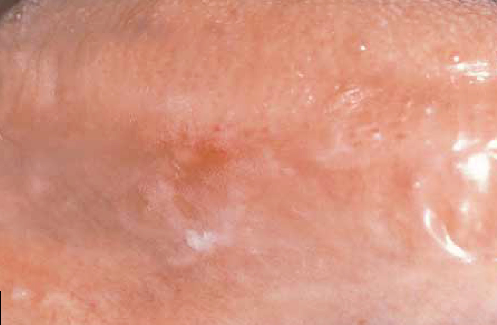
Speckled leukoplakia have a high risk of being premalignant – 23.4%

of the buccal mucosa showed moderate epithelial dysplasia.
Erythroplakia lesions – red patches
More likely to be dysplastic than Leukoplakia.
In Erythroleukoplakia, the red patches are more likely to be dysplastic and thus make sure a biopsy includes these parts of tissue!
More in older men
High risk sites:
- Floor of mouth
- Lateral tongue
- Retromolar pad
- Soft palate
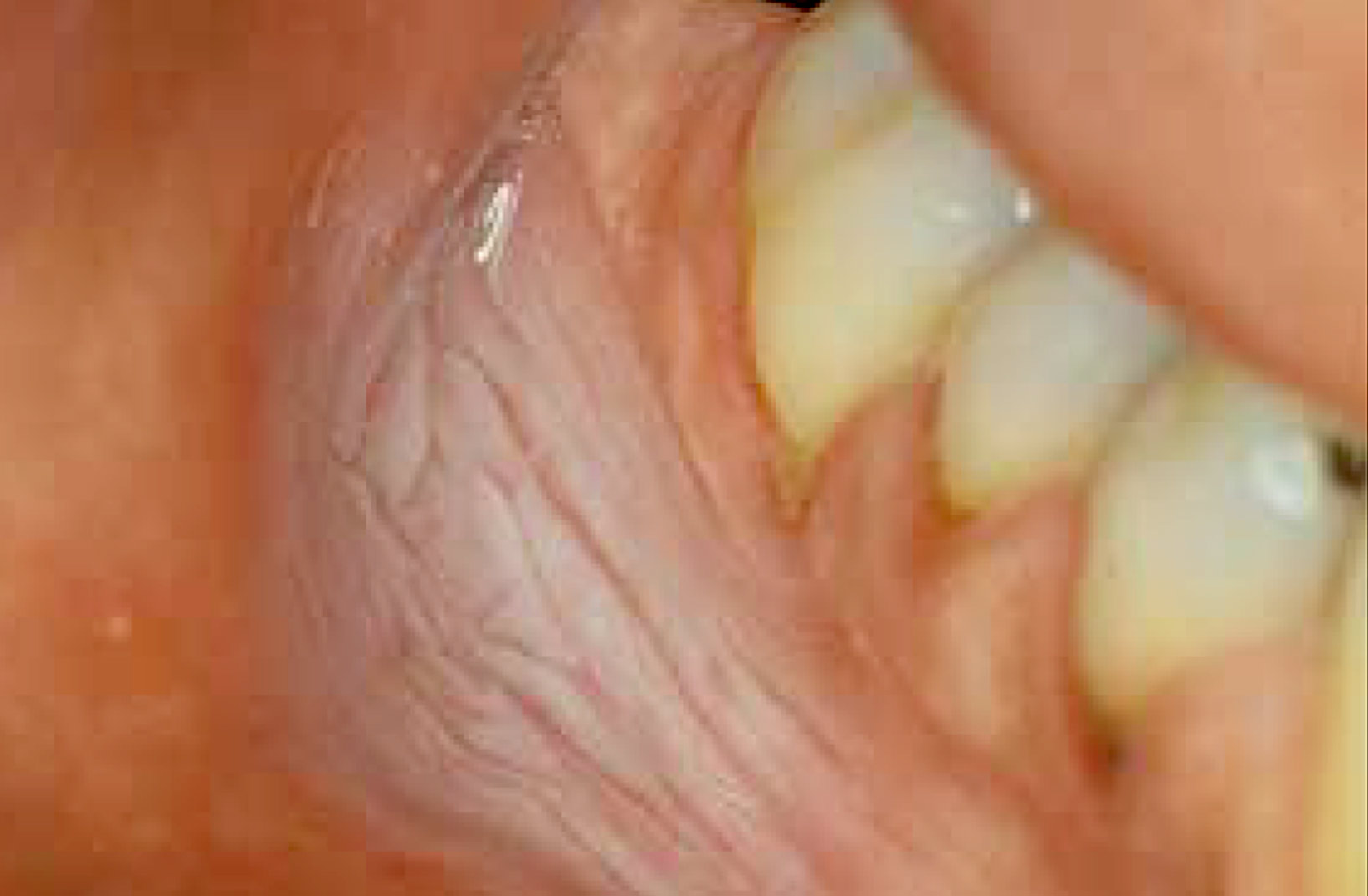
lateral border of the tongue showed carcinoma in situ on biopsy.
Tobacco pouch keratosis
Buccal mucosa usually affected from holding smokeless tobacco or betel quid. Usually resolves after 4-6 weeks of ceasing the habit. Sometimes persisting lesions become dysplastic and become malignant.
of the mucosa in the mandibular buccal vestibule secondary to the
use of chewing tobacco.
Squamous cell carcinoma:
Early presentation is either as a Leukoplakia, Erythroplakia or Erythroleukoplakia. With time, the lesion becomes ulcerated and may become an exophytic mass with a fungal or papillated surface.
Others may become endophytic with depressed cores and raised/rolled margins. They may or may not be symptomatic.
Most oral cancers occur on the tongue followed by the floor of the mouth. Other sites are less common.


During a dental exam, make sure that all high risk sites (including lateral areas of the tongue) are carefully checked.
References including images
Neville, B. W., & Day, T. a. (2002). Oral cancer and precancerous lesions. Ca-A Cancer Journal for Clinicians, 52(4), 195–215. https://doi.org/10.3322/canjclin.52.4.195